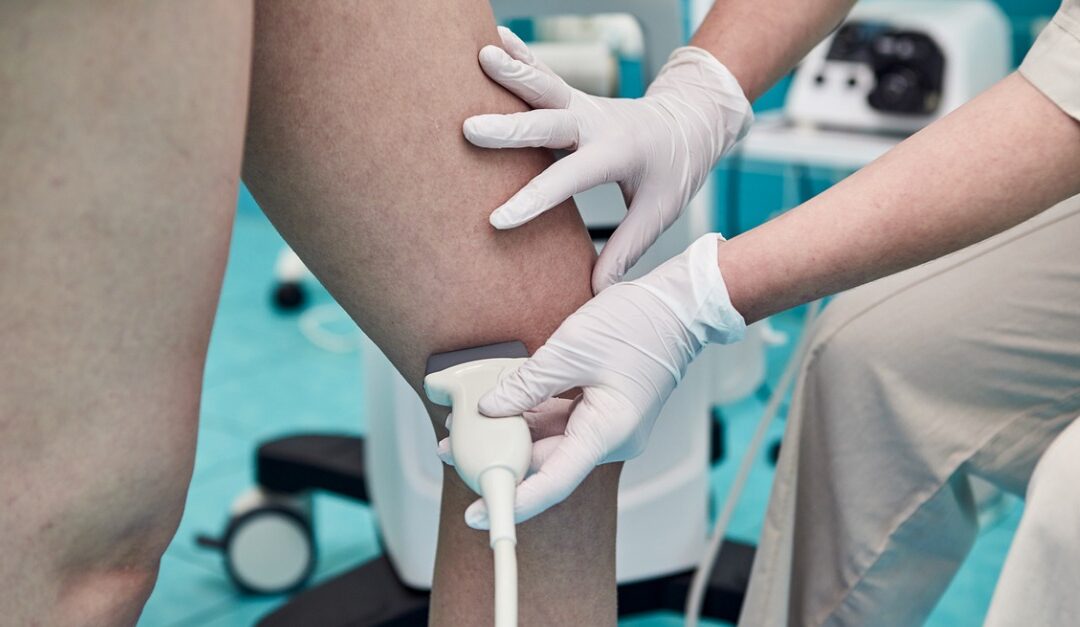
Venous ultrasounds are used to search for and diagnose certain conditions, including deep venous thrombosis, or blood clots. This imaging can also be used to assess the venous system for the presence of varicose veins and venous reflux disease.
Ultrasound technology uses sound waves to create images of the veins and is fairly quick and non-invasive. If you’ve been scheduled for a venous ultrasound at our facility, here’s what to expect from your appointment.
How Is a Venous Ultrasound Performed?
As with other types of ultrasounds, you’ll start by lying on an examination table that can be moved as needed. The technologist may have you stand depending on the assessment needed. From there a clear gel will be applied to the area of examination. The gel is a conductive medium that helps form a bond between your skin and the ultrasound transducer. It helps to eliminate pockets of air, thereby allowing soundwaves from the transducer to pass into the body. Although it has a sticky consistency, it wipes off easily without leaving a residue behind.
During the exam, the technologist will move the transducer over areas of interest to capture images. They may use different angles or ask you to move into different positions to access different areas of the venous system. Typically, venous ultrasounds take 30 to 45 minutes, but may go longer depending on the exam’s complexity.
How Should You Prepare for a Venous Ultrasound?
Preparation for an ultrasound depends on the area being examined. If you’re having veins in the abdominal region examined, you may be asked to modify your food or water intake leading up to the appointment. Otherwise, venous ultrasounds typically require no preparation before the appointment. Prior to the exam, you may be asked to change into a gown and to remove any jewelry or clothing in the area being examined.
What Can You Expect During & After a Venous Ultrasound?
While the ultrasound technologist moves the transducer over your skin, you may feel some mild pressure. Typically, there’s no discomfort involved, but you may feel minor pain if they’re taking images over a particularly tender area. If a Doppler ultrasound exam is also being performed, there may also be various sounds that change with your blood flow.
While regular ultrasounds can produce images of the venous systems, they can’t show blood flow. But Doppler ultrasounds bounce high-frequency sound waves off red blood cells as they circulate to show how blood is moving. This can be helpful for diagnosing blocked or bulging arteries, decreased circulation, poorly functioning valves, and narrowing of arteries.
After your venous ultrasound is complete, you’ll be able to resume your normal routine right away. A radiologist will interpret the results, and one of our providers will discuss findings with you to determine a treatment plan, if needed.
For any known or suspected venous conditions, turn to Vascular Surgical Associates for the highest level of care and treatment. Our accredited lab features advanced venous imaging technology, and our highly trained staff will help you feel at ease during your exam. Find out more about our venous imaging or schedule an appointment by calling (770) 423-0595.


