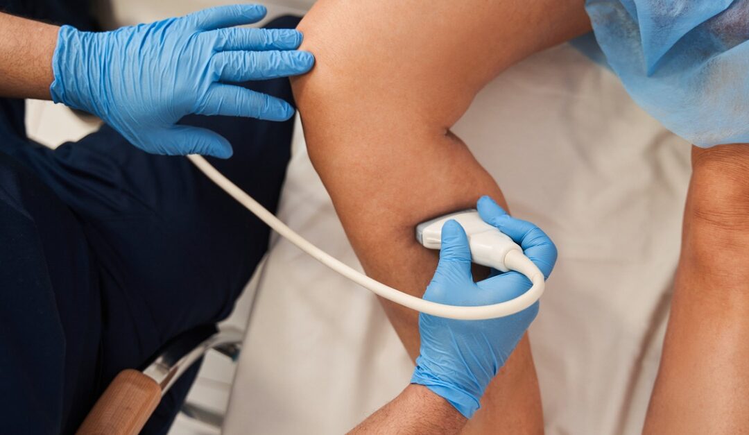
Vascular imaging allows doctors to use advanced technology for diagnosing and assessing conditions of your blood vessels. In some cases, medical imaging may also assist in minimally invasive procedures, equipping providers to perform procedures with less risk, reduced pain, and shorter recovery times, compared to traditional surgery.
There are many important applications for imaging in vascular care. Here’s a look into just a few of the most common ways vascular imaging is utilized.
Duplex Ultrasound Technology
A duplex ultrasound combines a traditional ultrasound — which targets sound waves to bounce from blood vessels then captures images — with Doppler ultrasound, which records sound waves to measure blood flow. This imaging can be used in the extremities to look for the presence of either deep or superficial venous thrombosis, as well as varicose veins and venous reflux disease. Duplex ultrasounds can also be used to assess blood vessels in the neck (including the carotid artery), as well as the abdomen.
In some cases, venous specialists may also use traditional ultrasounds to perform vein mapping, which creates a “roadmap” of your veins to guide medical procedures.
Computed Tomography Angiography (CTA)
Computed tomography angiography (CTA) blends traditional CT scans, which are a type of X ray that creates cross-sectional images using a computer, with a specially-formulated dye in order to create pictures of blood vessels. Also known as contrast material, the dye is injected through an intravenous line. This liquid material allows blood vessels to be seen with ease during the imaging.
CTA is widely used in vascular care to look for a range of conditions. For example, it may help identify an aneurysm, or give a clear view of vessels affected by atherosclerosis, which is narrowing caused by fatty deposits in the artery walls. Doctors may also use CTA to determine if your blood vessels have been damaged by an injury, or to look for blood clots that originally formed in your leg but may have traveled into your lungs.
Magnetic Resonance Angiogram (MRA)
A magnetic resonance angiogram (MRA) utilizes magnetic resonance imaging, or MRI. Whereas traditional angiography involves the placement of a catheter, MRA is far less invasive. This technology uses magnetic fields and radio waves to capture images of the blood vessels, as well as a contrast dye.
“An MRA is very helpful in many cases,” explains Dr. Lalani, “including investigating for an aneurysm, signs of a stroke, atherosclerosis, heart disease, aortic coarctation, or aortic dissection.”
Which Imaging Test Will You Receive?
Doctors base their decisions for which imaging type to use on a number of factors, including the patient’s symptoms and health history. For example, the dye in MRA testing involves gadolinium-based contrast, which is less likely to cause an allergic reaction in patients who may experience an issue with the iodine-based contrast used in CTA. But if your doctor needs a closer look at calcium deposits in the blood vessels, a CTA may be necessary, as MRA cannot detect those.
Whichever imaging test you may require, at Vascular Surgical Associates, our accredited vascular lab has advanced imaging technology, along with highly trained staff to diagnose and assess venous conditions with precision. Request a consultation with us through our website, or call (770) 423-0595.


